We are able to see the objects through the reflection, refraction of light. The human eye has a natural lens which allows the light to enter and the images to form. Like a regular pair of spectacles, our eyes also use a lens to adjust the focus and produce clear images. But have you ever wondered how this process occurs? Let’s find out:
Structure of the Human Eye:
Terminology used:
Sclera, Choroid, Cornea, Ciliary Body, Iris, Aqueous Humour, Crystalline Lens, Crystalline lens, Retina, Optic nerve, Fovea, Blind Spot, Vitreous Humour
Nearly 2.3 cm in diameter, the human eye is spherical in shape and is composed of a lens, adjustable shutter, photographic lens, and millions of light-sensitive cells to process the captured image. It's safe to say that it is almost like a camera.
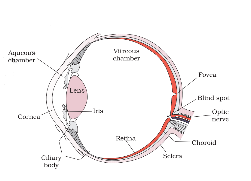
The Sclera, Choroid and Cornea form the outermost layer in the structure of the human eye. The light enters through the cornea which is in the form of a bulge at the anterior part of the eye. Sclera, composed of dense connective tissue is present behind the eye. Choroid forms a middle layer containing blood vessels and thickens to form a ciliary body at the posterior. The ciliary body continues to form a pigmented and opaque structure: iris. It is a dark muscular diaphragm and with the movement of the ciliary muscles, it controls the amount of light entering the eye.
When you enter a dimly lit room, your eyes take time to adjust and focus on the objects. The iris with the help of ciliary muscles relaxes the pupil so that more light can enter and vision becomes clear. Exact opposite happens if you move to a bright place. This time the iris contracts the pupil and lesser amount of light enters.
Aqueous Humour , a thin and watery fluid peasant between the cornea and the lens, not only nourishes the lens but also maintains its shape.

From the pupil light enters and falls on the crystalline lens. The lens focuses the light rays so that a clear, real, inverted image is obtained on the inner layer: retina. All the light-sensitive cells [Ganglion cells, Bipolar cells and Photoreceptor cells] on the retina are activated and the image is sent through the optic nerve to the brain for processing. The brain inverts the image and we can see the world as it is.
In the retina, photoreceptor cells rods (respond to brightness, dullness) and cones (respond to red, blue and green light) are present. Fovea is a point in the retina that is densely packed with cones while the Blind Spot in the retina lacks either of the photoreceptor cells. Vitreous chamber filled with transparent gel vitreous humour is present between lens and retina to maintain shape and pressure in the eyeball.
“Eyes are windows to the soul.”

With the integration of e-learning, e-books etc, we spend our time glued to device screens from a very young age. The first step in eye-care would be to divide your screen time in a day and include creative forms of entertainment like arts and crafts, reading, solving puzzles, indoor games (ludo, chess etc). Maintaining a proper sleep schedule of at least 7 to 8 hours. Washing your eyes with normal to cool water daily and taking a healthy diet rich in Vitamin A can make your eye-sight strong even till old age.